Retinal Imaging Makes Eye Exams Easier and More Comfortable Than Ever Before
|
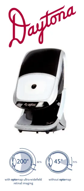
Eye exams should be among the easier exams that you need to get, but the tests where they dilate your eyes and leave you hiding behind those flexible film "sunglasses" can be enough to make anyone avoid the optometrist. For those of you who have dreaded eye checkups because of this, there's a new game in town called digital retinal imaging. This is essentially a photograph taken of the inside of your eye, allowing you and the eye doctor to both see your retina and associated structures. Because this imaging doesn't require dilation or other uncomfortable procedures, it is a great addition to your eye health exam. A Special Type of PhotographyDigital Retinal Imaging gives you and your optometrist a clear picture of what your retina and macula look like. It allows your eye doctor to better examine for eye disease that can lead to permanent vision loss. This includes ruling out glaucoma, macular degeneration, retinal detachments and many other sight-threatening conditions. The advantages of this imaging are four-fold. First, is the convenience as it does not require eye drops or your pupils to be dilated. Another is the fact that the resulting picture is large and clear, giving a 200 degree view of the inside of your eye. A third is that both you and your doctor get to see the picture at the same time, allowing the two of you to discuss exactly what's going on. The fourth may be the most important. The imaging gives the doctor a record that can be saved and used as a comparison the next time you have the imaging done. |
|
Technology at Epic Vision Eye CentersAll Epic Vision Eye Centers are proud to have cutting edge retinal imaging technology in the form of Optos Daytona Optomaps. The Optos Daytona allows us to see retinal and optic nerve structures in ways not visible through regular exam methods. The Daytona Optopmap uses light to provide a high resolution scan meant to pick up early signs of structural change or disease. It allows us to see the smallest changes in the retina and optic nerve with great accuracy and high definition, making early and accurate detection of common sight threatening eye conditions such as glaucoma, macular degeneration and diabetic retinopathy, easier than ever before. | |